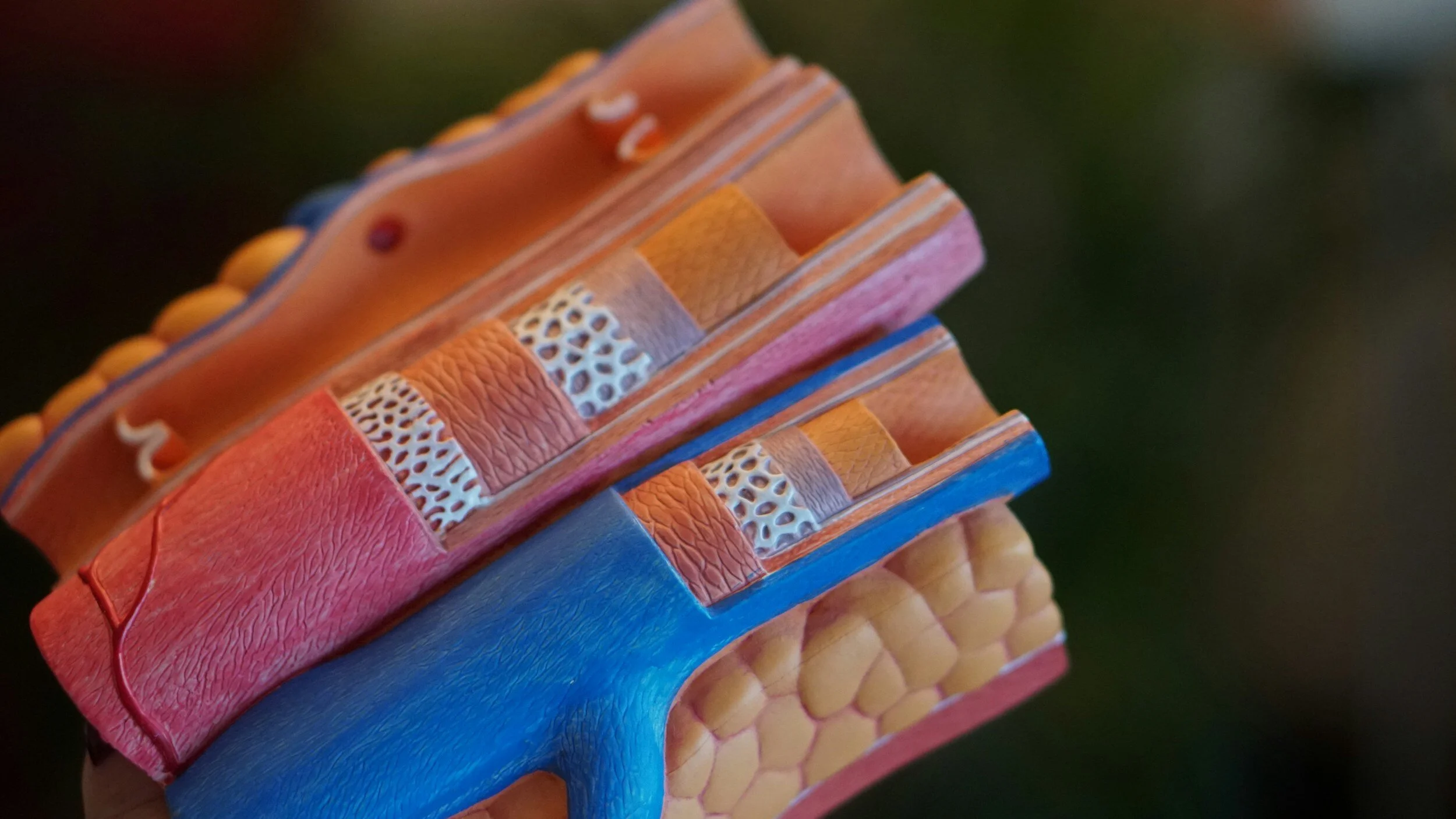
BP and Non-Invasive Vascular Assessments
We provide vascular CVD risk monitoring by measuring, central and not just peripheral arterial stiffness. Vascular health monitoring — central (aortic) BP, arterial stiffness (AIx), 24-hour ambulatory BP (Oscar 2™ with SphygmoCor® Inside) and Lumify ultrasound adjunct (PWV, PSV/EDV, RI).
Non-invasive central pressure and arterial stiffness assessment & 24-hour BP with central haemodynamics

-
What we measure (non-invasive):
Central (aortic) BP & arterial stiffness (e.g., Augmentation Index—AIx) via SphygmoCor® cuff/sensor.
24-hour ABPM (Oscar 2™ with SphygmoCor® Inside) for day/night pattern, BP load and central pressure estimates.
Optional “active” response: repeat measures after a gentle step/walk to see how central BP/AIx behave under light load.
Ultrasound adjunct (Lumify): focused vascular ultrasound to support PWV estimation (transit-time) and arterial wall assessments (e.g., carotid diameter/IMT**)** where appropriate.
Why it matters: Central pressure and stiffness reflect the load on the heart and the pulse reaching pressure-sensitive organs (e.g., kidneys). Findings guide exercise intensity, BP targets, and follow-up.
Scope: Functional assessment to support care — non-diagnostic; abnormal findings referred to GP/cardiology.
-
Clinic (resting / active) – central haemodynamics
Central (aortic) SBP/DBP/PP
Augmentation Index (AIx / AIx@75) — wave reflection / stiffness context
(If enabled)Pulse Wave Velocity (PWV) — transit-time estimate
“Active” response: change in central BP / AIx after gentle step/walk (3–5 min)
24-hour ABPM (Oscar 2™ with SphygmoCor® Inside)
Day/night mean SBP/DBP, dipping status, 24-h BP load (% time above target), central pressure estimates, pulse pressure
Ultrasound adjunct (Lumify) – focused vascular Doppler(used selectively)
PSV(Peak Systolic Velocity) and EDV(End-Diastolic Velocity) from short, angle-corrected Doppler samples (e.g., carotid / common femoral)
RI (Resistive Index) = (PSV−EDV)/PSV(PSV−EDV)/PSV — simple resistance/afterload marker
Diameter / IMT snapshot(where image quality allows) — arterial size/wall context
How we use these: values are supportive/contextual alongside central BP/AIx and ABPM, and reported with acquisition notes (site, angle correction, sample gate, beats averaged). We emphasise serial change under similar conditions, not single absolute cut-offs.
How we read it (all modalities)
Prioritise patterns and trends: non-dipping/night-time elevation, high central PP, increased AIx or PWV, RI/PI shifts with training or medication timing, and the “active” response.
Translate to exercise prescription (starting intensities, HR/BP ceilings, recovery guidance) and practical medication-timing conversations with the treating clinician.
Caveats: caffeine, stress, recent exertion, hydration, and angle error affect readings; repeat under similar conditions for longitudinal validity. Ultrasound metrics depend on operator technique and are reported as adjunctive, not diagnostic.
SphygmoCor® Technology
Is used clinically for central arterial pressure waveform analysis (PWV and PWA) to better inform blood pressure and cardiorenal management.Use it to check central carotid PWV and compare both Ultrasound and SphygmoCor for absolute piece of mind about relative risks. We send images of the findings on request.
We note that the site of measurement in central BP is important; a meta-analysis has found a difference in the Relative Risk of CVD endpoints if central pulse waveform recording site is Carotid artery (n= 498) 1.48 (CI: 0.86, 2.55)or Radial artery(n= 48561) 1.07 (CI; 1.03, 1.11). - (Li et al., 2024).
CVD endpoint = cardiovascular mortality, myocardial infarction, stroke, heart failure, and re-vascularizations (coronary artery bypass graft surgery or percutaneous coronary interventions.The SphygmoCor system allows physiologists to individualise care for patients with hypertension, renal disease, COPD, diabetes, and heart failure, & help physicians tailor medication dosages and monitoring vascular changes following treatments (2000+ per reviewed papers).It also measures central BP variables and Mean Arterial Pressure (MAP) along with the central stiffness variables Pulse Wave Velocity (PWV) and the peripheral Augmentation Index (Alx) standardised to a heart rate of 75 beats and the subendocardial viability ratio (SEVR), also known as the Buckberg index, which is arterial stiffness parameter correlated with coronary flow reserve which makes it a useful parameter in assessing coronary microvascular circulation SEVR estimates it is a measure of the balance between cardiac blood flow supply and demand.
-
Who benefits:
Hypertension or borderline BP; masked/white-coat patterns; exercise prescription in cardiac/metabolic risk; kidney risk; older adults; insurer/employer baselines.
When ABPM adds value: Suspected masked/white-coat HTN; assessing medication dose & timing effects (e.g., morning surges, evening troughs); night-time non-dipping; variable clinic readings.
Considerations: Arrhythmias can affect oscillometric accuracy; ensure correct cuff size; defer during acute illness or post-caffeine; interpret alongside symptoms and diary.
-
Resting central BP/AIx (≈30 min):
Quiet rest 5–10 min → cuff + tonometry/oscillometry captures → quality index checked.
“Active” response (≈45 min total):
Resting capture → gentle step/walk → repeat capture within 3–5 min.
24-hour ABPM (fit 15 min, return next day):
Fit cuff/recorder; diary for sleep/activity; inflations every ~20–30 min by day, less at night.
Preparation: Avoid caffeine for 3 h before clinic visit; wear short-sleeved top; take usual meds unless your doctor advises otherwise. ABPM: loose sleeve, plan brief shower/refit.
-
Deliverable:
Clinician PDF in 2–3 business days (priority 24 h). Patient receives plain-English summary.
Data:PDF is primary; CSV available on request for ABPM & AIx; ECG raw (SCP-ECG/XML) available from ECG services if relevant.
Identifiers & security: Coded filenames (no PHI); encrypted at rest/in transit; retention 7 years.
Cross-links: We align vascular results with exercise prescription (HR/BP limits) and, where relevant, with biomarkers or BIA.
-
Noninvasive central blood pressure assessments individualise treatment decisions and help get patients to their health goals faster.
Hypertension management using central BP can produce similar outcomes to therapeutic intervention but with less medication required.[4][5]
Antihypertensive medications have differential effects on central vs. brachial blood pressure, which can explain variability in clinical outcomes.[6]
Central blood pressure is more predictive of cardiovascular (CV) outcomes than brachial blood pressure, primarily due to proximity to target organs.[7]
(Augmentation Pressure: Central systolic and pulse pressures are increased in the presence of stiffer arteries [1][2][3], causing a higher ventricular after-load and energy used by the heart to pump blood around the circulatory system. Evaluation of the central pressure waveform contour allows for determination of augmentation pressure (AP) and the augmentation index (AIx), two parameters that quantify the increased pressure.)
-
A better predictor of outcome: A multi-year, NIH study in more than 3,500 high risk subjects demonstrated that central pulse pressure was more strongly predictive of CV events. Specifically, central pulse pressures > 50mmHG had a 20% increased risk of having a CV event in the next 5 years. Brachial pulse pressure did not show a similar threshold.[7].
24-hour Central Blood Pressure: Oscar 2 with SphygmoCor combines the superior predictive power of Central Blood Pressure with the established standard for hypertension diagnosis providing a 24-hour study with up to 250 brachial and central blood pressure measurements.
For Cardiac Rehabilitation it is noted by Aslanger er al., 2017 that some measures may be needed to be undertaken before CR to optimise exercise induced improvement adjusting baseline supply–demand ratio. These may include reducing afterload by decreasing peripheral vascular resistance and wave reflections (e.g., peripheral vasodilators) or augmenting diastolic blood flow by increasing duration of the diastole (e.g., ivabradine).
-
O’Rourke et al. Br J Clin Pharmacol 2001;51:507-22
Sharman JE et al., J Hum Hypertens 2008 (12):838-44
McEniery et al., Hypertension 2008 6(51):1476-82
Sharman et al., Hypertension. 2013 62:1138-45
Kosmala et al., Am J Hypertens. 2015 108
Williams B et al., Circulation 2006 113: 1213-25
Roman, et al., JACC 2009 54:1730-4
London GM, Hypertension 2001 38:434-8
Aslanger, E., et al., 2017 Anatolian journal of cardiology, 17(1), 37-43