
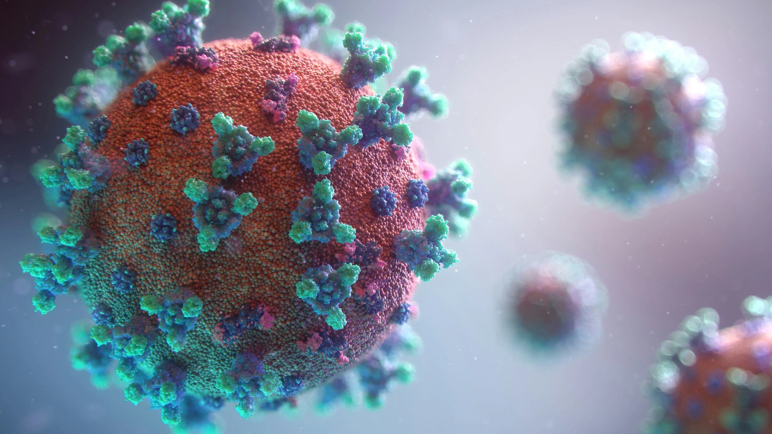
Long COVID, ME/CFS and PoTS. Measured, not guessed.
Ask Yourself, Could This Be You ?
Usually worse on standing, in heat, after meals, and in the hours or days after activity.
Light-headedness on standing, or near-fainting
Racing or pounding heartbeat when upright
Exhaustion out of proportion to what you did
A crash a day or two after activity, not during it
Brain fog and difficulty concentrating
Breathlessness or chest discomfort
Headaches, nausea, shakiness, cold or clammy hands
Worse in hot weather or a hot shower
Worse after skipping a meal or on a low-salt diet
Cannot stand still for long, but better walking
Training that used to be easy is now impossible
Sleep that does not refresh you
What to Expect
If every test has come back normal, it may be because the abnormality only appears when you stand up, or in the minutes after you exert yourself. Nobody has looked there yet. We do.
Long COVID and ME/CFS share almost the same picture
Post-viral onset after Epstein-Barr and glandular fever has been recognised for decades. COVID-19 produced the same pattern at scale.
❋ Why the standard workup comes back normal
Most people who come to us have already had the bloods, the ECG, often an echocardiogram. Everything was normal, and they were sent away with a label of anxiety, deconditioning, or nothing at all.
That is not anybody's fault. A resting ECG measures the heart lying still. A cuff on a seated arm gives one number, twice. Neither can detect a circulation that only fails after a person stands up, or only fails in the minutes after they have exerted themselves.
That is where the abnormality lives in long COVID and in ME/CFS. It is why the tests keep coming back clear, and it is exactly what our assessment is built to provoke and measure.
❋ An overlapping structure
The overlap is not a coincidence and it is not controversial. A large proportion of people with long COVID meet the established diagnostic criteria for ME/CFS, and the defining feature is the same in both: post-exertional malaise, a delayed worsening that arrives a day or two after activity rather than during it.
The physiology is also the same. An early metabolic threshold, the body switches to an unsustainable fuel system at a much lower workload than expected. Impaired autonomic recovery after exertion. And, very often, a heart that is not filling adequately when upright, which limits what it can deliver regardless of how fit the person is.
What Makes Our Tests Different
Standard orthostatic testing asks you to stand up from rest. That is the right first test and it identifies many people.
We can also apply the orthostatic challenge to a circulation that has already been destabilised by a sub-maximal Micro-Exertion Autonomic (MEA) challenge. For a substantial group, a stand from rest is not a strong enough provocation, and the problem only appears when the two are combined.
Throughout, we measure central aortic pressures and beat-by-beat pulse contour, using internationally validated vascular testing equipment and simultaneous ECG, not just cuff readings. Blood pressure can look perfectly normal while the volume the heart ejects with each beat is falling steadily. A cuff cannot see / read this.
-
Long COVID is increasingly recognised as a multi-system condition. In some people, symptoms appear to involve abnormal regulation of the circulation, autonomic nervous system, skeletal muscle and cellular energy production.
One emerging research model, developed by Prof. Klaus Wirth and colleagues, proposes that abnormal control of small blood vessels may contribute to an imbalance between vasoconstriction and vasodilation.
-
↓
Abnormal distribution of blood flow
Some tissues may become relatively under-perfused, while abnormal microvascular flow or shunting may allow excessive flow through other vascular pathways without normal oxygen extraction.
↓
Muscle and brain oxygen delivery may become inadequate during stress
↓
Earlier anaerobic metabolism and muscle acidosis
↓
Altered sodium and calcium handling within muscle cells
↓
Mitochondrial stress and reduced energy availability
↓
-
Post-viral disturbance / autonomic dysfunction
↓
Altered vascular signalling
β2-adrenergic signalling ↓ —reduced vasodilation and altered muscle ion regulation
α-adrenergic sympathetic activity ↑ — excessive vasoconstrictor drive in some patients
Endothelial / muscarinic signalling may be impaired
Small-fibre and other microvascular abnormalities may further affect vascular regulation
What Does Med-Ex Measure, There Are 3 Steps?
Rather than relying on a single test, Long Covid Phenotyping Tests examine how several physiological systems behave together.
1. Autonomic & Orthostatic Phenotyping
Examines cardiovascular responses to standing and gravitational stress using:
Active stand testing
Head-Up Tilt Table testing where indicated
Continuous blood-pressure and heart-rate monitoring
ECG and heart-rate variability
Baroreflex and autonomic response assessment
Valsalva testing where clinically appropriate
Looks for: POTS-type responses, orthostatic hypotension, inappropriate sympathetic activation, abnormal heart-rate responses and impaired cardiovascular recovery.
2. Exertional & Vascular Phenotyping
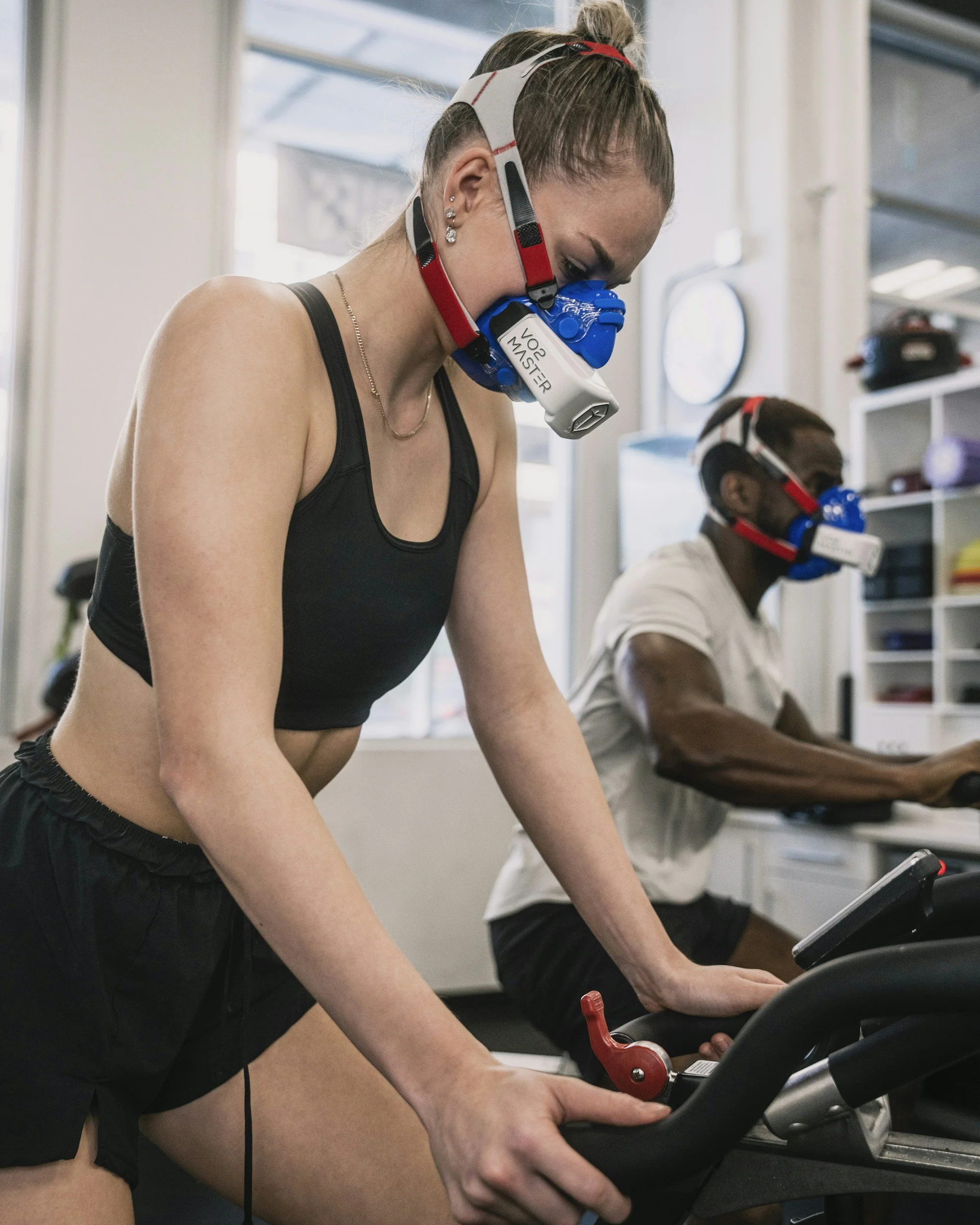
For patients able to tolerate it, a brief, carefully controlled low-exertion challenge can be used rather than automatically performing a maximal exercise test.
Testing may include:
Resting central blood pressure and Pulse Wave Analysis
Pulse Wave Velocity
ECG and heart-rate response
A brief sub-maximal MEA challenge
Immediate and serial recovery measurements
Central pressure and Subendocardial Viability Ratio response
Heart-rate recovery and autonomic recovery
The exertional component can be reduced or omitted in patients at significant risk of Post-Exertional Malaise (PEM).
Looks for: disproportionate cardiovascular stress, abnormal recovery, chronotropic abnormalities and changes in central haemodynamics following a very small physiological challenge.
3. Comprehensive Long COVID Phenotyping
Combines autonomic, orthostatic and vascular assessment with selected laboratory markers according to the patient's clinical presentation. These can be processed as in-clinic screening analysis or arranged through Awanui Labs where clinically indicated, may include
Potential markers include:
IL-6
CRP / Serum Amyloid A
D-dimer
NT-proBNP
Cardiac troponin
Cystatin-C
Creatinine and electrolytes
Cortisol
Renin, noradrenalin, aldosterone
The objective is not simply to confirm that symptoms exist, but to identify which physiological systems appear most disturbed in the individual patient and provide objective findings that can assist subsequent clinical management.
Phenotyping — not a one-size-fits-all test
Two people with Long COVID may experience similar fatigue while demonstrating very different physiological patterns.
One may predominantly demonstrate orthostatic intolerance, another excessive sympathetic cardiovascular activation, another an abnormal vascular response to exertion, and another a stronger inflammatory or metabolic component.
Med-Ex Long Covid Phenotyping Tests are designed to identify these differences.
Why the answer matters: two conditions, two opposite treatments
This is the single thing testing is for. No consultation, questionnaire or history can reliably tell these two apart, because they describe themselves the same way. A measurement can.
If the picture is PoTS, without post-exertional crashes
Structured reconditioning works. A progressive programme that begins recumbent or semi-recumbent and advances slowly, always below your measured threshold.
In the trial that established this approach, published by Fu and Levine in Hypertension in 2011, a majority of patients no longer met the criteria for PoTS after three months, with increased blood volume, increased cardiac mass and a lower standing heart rate.
Known as the Levine or CHOP protocol. It depends entirely on knowing where your threshold actually sits.
If post-exertional malaise is present - with energy crashes
Progressive exercise is the wrong treatment, and pushing through makes things worse. The approach here is energy management: staying inside your envelope, and expanding it only as the envelope itself changes.
NICE guideline NG206, published in 2021, no longer recommends graded exercise therapy for ME/CFS and long COVID and it places energy management at the centre of care. This is in order to protect muscle energy metabolism.
This is where a measured pacing ceiling earns its keep, because the whole approach depends on a number.
The detailed mechanisms of the condition defined here:
What applies either way
A pacing ceiling that is measured, not estimated. Age-predicted maximum is commonly 15 to 20 bpm away from a person's real maximum, and heart rate zones calculated from it can be wrong by a wide margin. In this group, an estimated ceiling fails the people who can least afford it.
Volume expansion. Salt and fluid loading, at levels agreed with your doctor and where there is no reason to avoid it. Inexpensive, not a prescription, and we can measure whether it worked by repeating the orthostatic test and your hydration levels using bioelectrical impedance analysis.
Compression. Abdominal compression reduces orthostatic tachycardia and symptoms in PoTS, published in JACC in 2021, with more recent work showing abdominal-only garments are effective. Blood pooling in the abdomen is the target, which is why waist-high garments outperform calf-length ones.
Finding the treatable things that get missed, iron deficiency above all, which contributes independently to both fatigue and orthostatic symptoms.
Information your doctor can prescribe from. Where medication is warranted, the choice differs depending on which physiological pattern you have. That decision belongs to your doctor, and the measurement is what lets it be made on data.
Objective documentation of capacity and postural tolerance for ACC, insurers, schools and employers, and a baseline to measure change against later.
For referrers
We accept referrals from general practice, sport and exercise medicine, cardiology, rheumatology, paediatrics, immunology and allied health, and are most often useful in four situations.
A post-viral fatigue patient who needs a pacing threshold. A measured ventilatory threshold rather than a percentage of predicted maximum. Adherence improves, because the number is theirs.
A patient whose active stand was equivocal. We can apply the orthostatic challenge after a bounded exercise provocation, with central haemodynamic monitoring throughout, and from October with continuous stroke volume on tilt.
A patient who needs functional capacity documented for ACC, an insurer, a school or an employer.
An athlete whose performance has dropped without explanation. Gas exchange, thresholds, central pressures and rhythm in a single session.