
Unmasking POTS, Long-COVID, and Dysautonomia
1. The Viral Antecedent: Where Dysautonomia Begins
Postural Orthostatic Tachycardia Syndrome (POTS), Long-COVID, and Myalgic Encephalomyelitis/Chronic Fatigue Syndrome (ME/CFS) rarely appear in a vacuum; they frequently follow an acute vi1ral infection.
Prospective cohort studies have established Epstein-Barr Virus (EBV) as a prospectively demonstrated trigger for post-infective fatigue syndromes [10]. The risk is particularly pronounced in younger populations, with data showing that 13% of adolescents meet ME/CFS criteria six months after acute infectious mononucleosis [11]. Prospective tracking of college students has further validated these long-term risks following mononucleosis [12].
Recent immunological investigations demonstrate that early EBV reactivation is a powerful predictor for anticipating post-acute COVID-19 sequelae (Long-COVID) [13]. Cohort studies continue to observe significant relationships between long-COVID prevalence and underlying EBV reactivation [14]. Understanding this viral foundation is the first step in the Med-Ex phenotyping process.
2. The Pathophysiology: Competing & Parallel Mechanisms of Failure
Standard medical models frequently struggle to explain severe symptoms like cognitive "brain fog" and post-exertional malaise (PEM). However, advanced physiological research provides two prominent, and potentially parallel, pathophysiological models.
The Autoantibody & Hypoperfusion Hypothesis 1
A unifying hypothesis proposed by Wirth and Scheibenbogen suggests these syndromes are driven by autoantibodies directed against beta-2-adrenergic receptors [7]. When these receptors are disabled, the cardiovascular system defaults to intense vasoconstriction and sympathetic nervous system overdrive [7]. To survive this severe vasoconstriction, hypoxic tissues release massive amounts of bradykinin, a powerful vasodilator that fundamentally breaks the body's fluid balance.
Table 1: Potential Effects of Bradykinin on Vascular Filling
-
Bradykinin drastically increases endothelial permeability, forcing intravascular fluid to leak out of the vessels and lowering total blood volume. This is exaggerated by dysfunctional beta-2 receptors. (Clinical marker: Elevated plasma albumin).
-
Bradykinin acts as a potent vasodilator in the kidneys, inhibiting sodium reabsorption in the distal tubule and acting like a mild diuretic, forcing the body to excrete necessary fluid. (Clinical marker: Decreased creatinine/urea).
-
Normally, low blood volume triggers the kidneys to produce Renin to retain fluid. The renal actions of bradykinin completely annihilate the low-pressure and low-sodium signals that the kidneys rely on, explaining the paradoxical failure of the RAAS system.
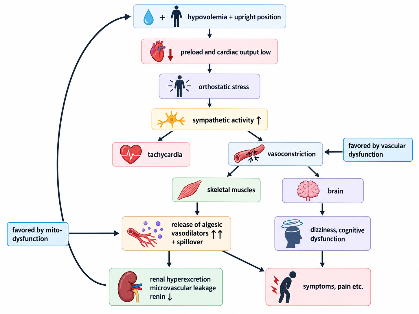
Figure 1: The Master Systemic Cascade (Wirth & Scheibenbogen, 2020). Autoantibody-induced receptor desensitisation leads to simultaneous vascular dysregulation, cerebral hypoperfusion, and sympathetic overdrive [7].
Furthermore, these beta-2-adrenergic receptors also govern the Na+/K+-ATPase pumps inside skeletal muscles [9]. When these pumps fail, it triggers a catastrophic intracellular sodium overload [9]. To clear the sodium, the cell imports massive amounts of calcium, which directly poisons the mitochondria [9]. This calcium-driven bioenergetic failure is the precise mechanism behind post-exertional malaise [9].
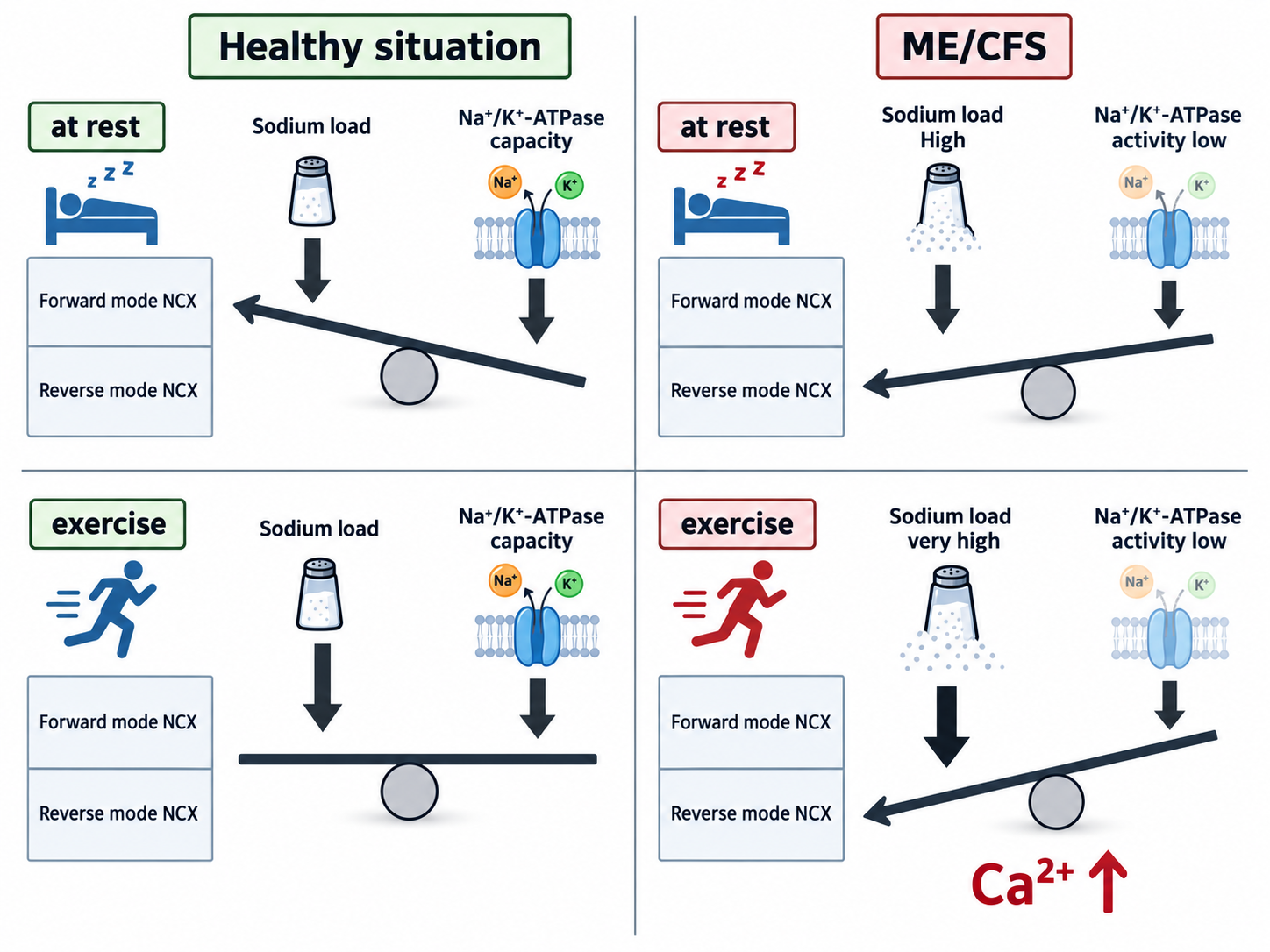
Figure 2: Skeletal Muscle and Mitochondrial Dysfunction (Wirth & Scheibenbogen, 2021). The failure of the Na+/K+-ATPase pump leads to intracellular sodium overload and subsequent mitochondrial calcium poisoning [9].
To clear the sodium, the cell imports massive amounts of calcium, which directly poisons the mitochondria [9]. This calcium-driven bioenergetic failure is the precise mechanism behind post-exertional malaise [9]. Furthermore, while ME/CFS often presents with severe neurological symptoms mimicking encephalitis, this model attributes these symptoms to disturbed neurovascular coupling and cerebral hypoperfusion, functionally starving the brain of blood [8].
The Intracranial Hypertension & CSF Hypothesis
Conversely, emerging research proposes a structural and fluid-dynamic mechanism, noting that typical symptoms of ME/CFS overlap considerably with those of idiopathic intracranial hypertension (IIH) [23]. Recent clinical studies demonstrate that a significant proportion of ME/CFS patients (up to 38% in a targeted cohort) exhibit elevated cerebrospinal fluid (CSF) opening pressures of 20 cmH2O or greater [23].
Researchers propose that elevated intracranial pressure (ICP) in this subgroup may be a consequence of impaired lymphatic and glymphatic clearance, leading to toxic buildup and neuroinflammation [23]. Alternatively, structural abnormalities in the craniocervical region may impede the natural flow of blood or CSF, altering central nervous system activity [23]. Notably, a substantial number of these patients experience significant symptomatic relief of brain fog and headache following CSF subtraction via lumbar puncture [23].
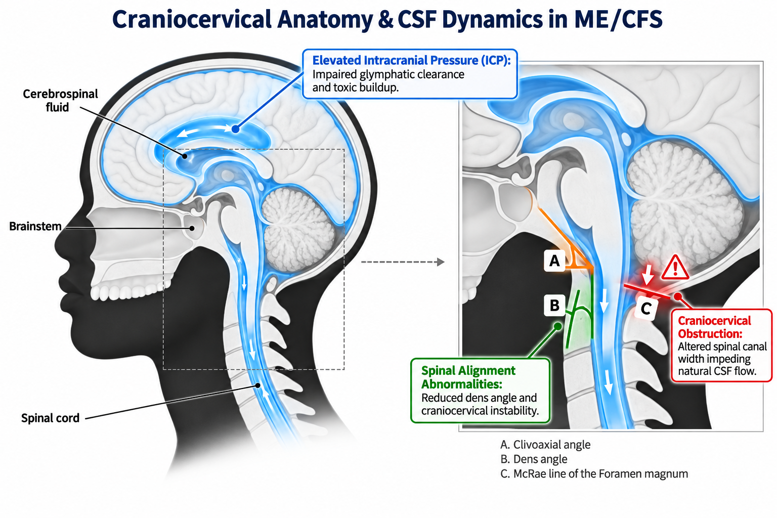
Figure 3: Craniocervical Anatomy and CSF Dynamics (Jolley et al., 2026). The transition area between the brain and cervical spine, illustrating the foramen magnum and structures that may influence CSF flow and contribute to elevated intracranial pressure in ME/CFS subgroups [23].
Furthermore, following 7-Tesla MRS scanning of the brain (pgACC and dACC) and calf muscle, alongside cognitive assessments, distinct metabolic differences emerged. Compared to controls, ME/CFS patients had elevated lactate in the areas of interest that controls fatigue, indicating energetic stress and mitochondrial dysfunction. Conversely, long COVID patients showed reduced total choline. Skeletal muscle metabolites did not significantly differ between the groups. The reduction in choline in long COVID is of particular interest given the recent association between blood clots and ‘brain fog,’ supported by animal studies showing choline might prevent intravascular coagulation [24].
3. The Hemodynamic Reality: What Standard Tests Miss
Standard resting tests (like a 12-lead ECG or static blood pressure cuff) are inherently incapable of diagnosing these dynamic conditions.
Preload Failure: Invasive cardiopulmonary exercise testing has definitively demonstrated that a primary mechanism in ME/CFS is preload failure, leading to severely impaired oxygen extraction at the muscular level [1].
Cerebral Hypoperfusion: During head-up tilt testing, patients with ME/CFS suffer from significant, measurable reductions in cerebral blood flow, or at the very least significant changes in the flow even in the absolute absence of systemic hypotension or tachycardia [2].
Long-COVID Similarity: This exact pattern of orthostatic symptoms and reductions in cerebral blood flow is identically mirrored in Long-COVID patients [3].
4. The Diagnostic Blind Spot: The Med-Ex Approach
At Med-Ex, we utilise advanced central haemodynamics and beat-to-beat autonomic monitoring to unmask the exact physiological bottleneck. Our proprietary diagnostic reports provide referring physicians with objective data that standard assessments miss:
Diagnostic Thresholds: We accurately map orthostatic responses against the 2019 NIH Expert Consensus criteria for POTS [4], carefully applying specialised thresholds, such as the ≥40 bpm adolescent criterion established in paediatric literature [5].
The Illusion of the Arm Cuff: A standard arm blood pressure cuff may show a widening pulse pressure upon standing, falsely suggesting a healthy circulatory response. However, our central aortic measurements routinely reveal the truth: the central pulse pressure is actually falling, unmasking severe venous pooling.
SEVR Decomposition: We decompose a Subendocardial Viability Ratio (SEVR) to prove whether coronary perfusion pressure is intact, demonstrating if the heart's struggle is driven by a catastrophic loss of diastolic filling time.
Structural & Craniocervical Exclusions: Our findings help guide essential secondary specialist referrals. For instance, detecting specific ectopic patterns warrants targeted echocardiography to exclude structural defects [6, 21, 22]. Likewise, identifying distinct encephalitic or pressure-related phenotypes can prompt referrals for upright craniocervical MRI to investigate foramen magnum or spinal canal abnormalities that may influence CSF pressure [23].
5. Evidence-Based Solutions and Treatment Pathways
Once testing isolates the specific mechanism, we provide targeted management pathways based on real-world data from large-scale patient cohorts [18]:
First-Line Physiological Interventions
Energy Management (Pacing): Where post-exertional malaise is present, graded exercise therapy is strictly contraindicated [19]. Instead, pacing and energy management score highest for patient efficacy (75.2%) [18]. We use our haemodynamic data to establish a precise maximum heart rate "ceiling" to protect cellular mitochondria.
Targeted Compression: Research confirms that abdominal-only or waist-high compression offers superior results by physically blocking splanchnic blood pooling [16, 17].
Volume Expansion: Aggressive sodium and fluid loading directly targets preload failure, offering significant relief for postural orthostatic symptoms [18].
Pharmacological & Structural Pathways (Specialist Directed)
Our data allows specialists to prescribe or investigate with physiological confidence:
Targeted Rate Control: Real-world outcomes indicate that specific rate-controlling agents (like ivabradine) often outperform standard beta-blockers, which can worsen symptoms in a subset of patients by removing necessary vascular compensations [15, 18].
Central Sympatholytics: Medications such as guanfacine show emerging clinical utility for cognitive deficits [20], but are clinically appropriate only when advanced testing confirms a central hyperadrenergic phenotype.
Neurological Investigation: If symptoms indicate potential intracranial hypertension, patients may be referred for specialised neurological assessment to evaluate CSF opening pressures and potential mechanical flow obstructions [23].
Clinical References
Joseph P, et al. Insights from invasive cardiopulmonary exercise testing of patients with myalgic encephalomyelitis / chronic fatigue syndrome. Chest 2021;160(2):642-651.
van Campen CLMC, et al. Cerebral blood flow is reduced in ME/CFS during head-up tilt testing even in the absence of hypotension or tachycardia. Clinical Neurophysiology Practice 2020;5:50-58.
van Campen CLMC, et al. Orthostatic symptoms and reductions in cerebral blood flow in long-haul COVID-19 patients. Medicina 2022;58(1):28.
Vernino S, et al. Postural orthostatic tachycardia syndrome: state of the science and clinical care from a 2019 NIH Expert Consensus Meeting, Part 1. Autonomic Neuroscience 2021;235:102828.
Singer W, et al. Postural tachycardia in children and adolescents: what is abnormal? Journal of Pediatrics 2012;160(2):222-226.
Binici Z, et al. Excessive supraventricular ectopic activity and increased risk of atrial fibrillation and stroke. Circulation 2010;121(17):1904-1911.
Wirth K, Scheibenbogen C. A unifying hypothesis of the pathophysiology of ME/CFS: recognitions from the finding of autoantibodies against beta2-adrenergic receptors. Autoimmunity Reviews 2020;19(6):102527.
Wirth KJ, et al. An attempt to explain the neurological symptoms of ME/CFS. Journal of Translational Medicine 2021;19(1):471.
Wirth KJ, Scheibenbogen C. Pathophysiology of skeletal muscle disturbances in ME/CFS. Journal of Translational Medicine 2021;19(1):162.
Hickie I, et al. Post-infective and chronic fatigue syndromes precipitated by viral and non-viral pathogens: prospective cohort study. BMJ 2006;333(7568):575.
Katz BZ, et al. Chronic fatigue syndrome after infectious mononucleosis in adolescents. Pediatrics 2009;124(1):189-193.
Jason LA, et al. Risks for developing ME/CFS in college students following infectious mononucleosis: a prospective cohort study. Clinical Infectious Diseases 2021;73(11):e3740-e3746.
Su Y, et al. Multiple early factors anticipate post-acute COVID-19 sequelae. Cell 2022;185(5):881-895.e20.
Gold JE, et al. Investigation of long COVID prevalence and its relationship to Epstein-Barr virus reactivation. Pathogens 2021;10(6):763.
Fu Q, et al. Exercise training versus propranolol in the treatment of the postural orthostatic tachycardia syndrome. Hypertension 2011;58(2):167-175.
Bourne KM, et al. Compression garment reduces orthostatic tachycardia and symptoms in patients with postural orthostatic tachycardia syndrome. Journal of the American College of Cardiology 2021;77(3):285-296.
Bourne KM, et al. Abdominal-only compression garments reduce orthostatic tachycardia and improve symptoms in patients with POTS. Canadian Journal of Cardiology 2026;42(6):1320-1328.
Eckey M, et al. Patient-reported treatment outcomes in ME/CFS and long COVID. PNAS 2025;122(28):e2426874122.
National Institute for Health and Care Excellence. Myalgic encephalomyelitis, or encephalopathy, chronic fatigue syndrome: diagnosis and management. NICE guideline NG206, 2021.
Fesharaki-Zadeh A, et al. Clinical experience with the alpha2A-adrenoceptor agonist guanfacine and N-acetylcysteine for the treatment of cognitive deficits in long COVID-19. Neuroimmunology Reports 2023;3:100154.
Silvestry FE, et al. Guidelines for the echocardiographic assessment of atrial septal defect and patent foramen ovale. Journal of the American Society of Echocardiography 2015;28(8):910-958.
Baumgartner H, et al. 2020 ESC guidelines for the management of adult congenital heart disease. European Heart Journal 2021;42(6):563-645.
Jolley C, Bragée B, Soinne L, Huhmar H, Billing H, Bertilson B, Sjogren P. Cerebrospinal fluid opening pressure in relation to symptomatology and craniocervical anatomy in patients with myalgic encephalomyelitis/chronic fatigue syndrome. Frontiers in Medicine 2026;13:1869714.
Godlewska, B.R., Sylvester, A.L., Emir, U.E. et al. Brain and muscle chemistry in myalgic encephalitis/chronic fatigue syndrome (ME/CFS) and long COVID: a 7T magnetic resonance spectroscopy study. Mol Psychiatry 2025; 30, 5215–5226.